


Coksartrose- It is osteoarthritis of the hip joint.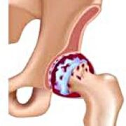 It is gradually developing, for several years, subject to progression, can be both a single factor and double.It is accompanied by the pain and the restriction of movements in the joint.In the last stages, the atrophy of the hip muscles and the shortening of the member are observed.The diagnosis is established on the basis of clinical symptoms and the results of radiography.In the first stages of coxarthrosis, conservative treatment.With the destruction of the joint, especially in youth and average age patients, surgery (endoprothetics) is indicated.
general information
Coksartrosis (osteoarthrosis or distorting osteoarthritis of hip joint) is a dystrophic degenerative disease.It generally develops at the age of 40 and over.This can be the result of various injuries and joint disease.Sometimes it occurs for no apparent reason.Coksartrose is characterized by a progressive progressive evolution.In the early stages, conservative treatment methods are used.In the subsequent stages, the joint function can only be restored.
In orthopedics and traumatology, coxarthrosis is one of the most common osteoarthritis.The high frequency of its development is due to a significant load on the hip joint and the generalized prevalence of congenital pathology - joint dysplasia.Women suffer from a little more often from coksartrosis than men.
The causes of coksartrose
Arthritis of the joint of the secondary joint (developed (developed as a result of other diseases) (developed as a result of other diseases).
Secondary coksartrosis can be the result of the following diseases:
- Joint hip dysplasia.
- Innate dislocation of the thigh.
- Losses.
- Aseptic necrosis of the thigh head.
- Infectious lesions and inflammatory processes (for example, hip joint articulation).
- Injuries (traumatic dislocations, hip neck fractures, pelvic fractures).
Coksartrose can be unilt or a double factor.With primary coxarthrosis, a concomitant lesion of the spine (osteochondrosis) and the knee joint (gonartrose) is often observed.
Risk factors
Among the factors that increase the probability of development of coxarthrosis include:
- Load increases constant on the seal.Most often observed in athletes in people with excess body weight.
- Circulatory disorders, hormonal changes, metabolic disorders.
- Pathology of the spine (cyphosis, scoliosis) or stop (flat feet).
- Elderly and senile age.
- A sedentary lifestyle.
Coksartrose itself is not inherited.However, certain characteristics (metabolic disorders, structural characteristics of the skeleton and weakness of the cartilage) can be inherited by the child's child.Consequently, in the presence of blood parents suffering from coxarthrosis, the probability of the occurrence of the disease is slightly increased.
Patanatomy
The hip joint is formed by two bones: iléon and femoral.The head of the thigh is articulated with the acetabulum of the iliac bone, forming a particular "hinge".During movements, acetabulum remains motionless and the femoral head moves in various directions, ensuring flexion, extension, abduction, productization and hips of rotation.
During the movements, the joint surfaces of the bones without hindrance compared to the others, thanks to the smooth, elastic and durable cartilage covering the cavity of the swivel cavity and the head of the thigh.In addition, the hyaline cartilage fulfills an absorbing function and is involved in the redistribution of the load during movement and walking.
In the joint cavity, there is a small amount of joint fluid, which plays the role of lubrication and provides nutrition of hyalin cartilage.The joint is surrounded by a dense and strong capsule.Above the capsule are large femoral and glued muscles, which provide movements in the joint and, with hyalin cartilage, are also shock absorbers which protect the articulation against injuries with unsuccessful movements.
With coxarthrosis, joint fluid becomes thicker and more viscous.The surface of dry hyalin cartilage loses softness, covered with cracks.Due to the roughness that appeared, the cartilage during the movements is constantly injured in each other, which causes their thinning and aggravates pathological changes in the joint.As coxarthrosis progresses, the bones are starting to deform, "adapt" to increased pressure.Metabolism in the joint deteriorates.In the last stages of coxarthrosis, severe atrophy of the muscles of the painful member is observed.
Coxarthrosis symptoms
The main symptoms of the disease include pain in joint, inguinal region, thigh and knee joint.In addition, with cokesartrose, stiffness of the movements and the rigidity of the joint, the disturbance of the walking, the lameness, the atrophy of the hip muscles and the shortening of the limb to the side of the lesion are observed.A characteristic of coksartrose is a restriction of the removal (for example, the patient is difficult when you try to sit on a chair).The presence of certain signs and their severity depends on the stage of coxarthrosis.The first and most constant symptom is pain.
HAS1st degree coksartrosisPatients complain of periodic pain, which occur after physical activity (running or walking extension).The pain is located in the joint, less often in the thigh or the knee.After rest, it generally disappears.The approach of 1st degree coxarthrosis is not broken, the movements are kept in full, there is no muscular atrophy.
On the X -ray of the patient with 1st degree coxarthrosis, light changes are determined: moderate unequal narrowing of the joint difference, as well as bone growth around the external or internal acetabulum edge in the absence of changes in the head of the femur.
HAS2 degree coksartroseThe pain becomes more intense, often appears at rest, radiates in the thigh and groin.After a significant physical activity, the patient with coksartrosis begins to be boxed.The volume of movements in the joint decreases: the abduction and internal rotation of the thigh are limited.
In x -ray images for 2nd degree coxarthrosis, a significant unequal narrowing of the joint deviation (more than half of the normal height) is determined.The femoral head is somewhat offset upwards, distorted and increased in size, and its contours become uneven.Bone growth with this degree of coxarthrosis appear not only on the interns, but also on the outer edge of the acetabulum and get out of the cartilage.
HAS3 -degree coksartroseThe pain becomes constant and concerned about patients not only during the day, but also at night.Walking is difficult during a movement, a patient with coksartrosis is forced to use a cane.The volume of movements in the joint is highly limited, the buttocks muscles, the hips and the lower legs are atrophied.The weakness of the muscles of the elimination of the thigh becomes the cause of the deviation of the basin in the front plan and shortening the member on the painful side.In order to compensate for the shortening, a patient with coksartrose, when walking, tilted the body towards the painful direction.For this reason, the center of gravity moves, the load on the painful joint increases sharply.
On 3rd degree coxarthrosis radiographs, a net narrowing of the joint gap, a pronounced expansion of the thigh head and multiple bone growths are detected.
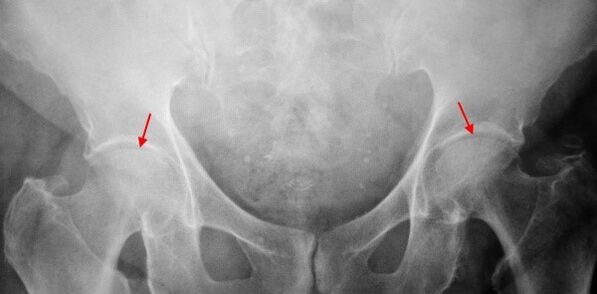
Diagnosis
The diagnosis of coxarthrosis is based on clinical signs and additional study data, the main one of which is radiography.In many cases, X -rays make it possible to establish not only the degree of coxarthrosis, but also the cause of its occurrence.Thus, for example, an increase in the angle of the coueticphysian, the scenes and the flattening of the acetabulum indicate dysplasia, and the changes in the form of the proximal part of the femur are indicated that coksartrose is a consequence of the disease of losses or young epiphysiolysis.On patients with patients with coxarthrosis, changes can also be detected indicating injuries.
Like other instrumental diagnostic methods of coxarthrosis, CT and MRI can be used.Computed tomography allows you to study in detail pathological changes by bone structures, and magnetic resonance imaging offers the possibility of assessing disorders by soft tissues.
Differential diagnosis
First of all, coxarthrosis must be differentiated from gonarthrosis (osteoarthrosis of the knee joint) and osteochondrosis of the spine.The atrophy of the muscles, which occurs at 2 and 3 stages of coxarthrosis, can cause pain in the knee joint, which are often expressed brighter than pain in the damage area.Consequently, with the patient's complaints concerning the knee pain, a clinic (inspection, palpation, determination of the volume of movements) is the study of the hip joint, and if coxarthrosis is suspected, to direct the patient to radiography.
Pain for radicular syndrome (compression of nerve roots) for osteochondrosis and some other spine diseases can imitate pain with coxarthrosis.Unlike Coksartrose, when pressing the roots, pain suddenly occurs, after a unsuccessful movement, a clear turn, lifting weights, etc., is located in the buttocks area and spreads along the thigh.A positive symptom of tension is detected - intense pain when the patient tries to lift a straightened member, lying on the back.At the same time, the patient freely takes his leg on the side, while in patients with coksartrose, the removal is limited.It should be kept in mind that osteochondrosis and coksartrose can be observed at the same time, therefore, in all cases, an in -depth examination of the patient is necessary.
In addition, cokesartrose is differentiated by trochanteritis (start -up scholarship) - aseptic inflammation in the field of glued muscles attachment.Unlike coxarthrosis, the disease develops rapidly, within 1 to 2 weeks, generally after a significant injury or physical activity.The intensity of the pain is higher than with coksartrosis.The limits of the movements and the shortening of the limb are not observed.
In some cases, with an atypical course of reactive disease or arthritis, symptoms resembling coxarthrosis can be observed.Unlike coxarthrosis, with these diseases, the peak of pain falls at night.Pain syndrome is very intense, can decrease when walking.Morning rigidity is characteristic, which occurs immediately after awakening and gradually disappears in a few hours.
Coxarthrosis treatment
The treatment of pathology is engaged in traumatician orthopedists.The choice of treatment methods depends on the symptoms and stage of the disease.At 1 and 2 stages of coxarthrosis, conservative therapy is carried out.During the exacerbation period of coxarthrosis, injection blocks, non-steroidal anti-inflammatory drugs (pyroxes, indomethacin, diclofenac, ibuprofen, etc.) are used.It should be kept in mind that the drugs in this group have not been recommended for a long time, as they can have a negative effect on the internal organs and remove the capacity of the hyalin cartilage to restore.
To restore damaged cartilage for coksartrosis, the funds of a group of chondroprotectors (chondroitine sulfate, cartilage extract, etc.) are used.To improve blood circulation and eliminate spasms from small vessels, vasodilating drugs (zinnarisine, nicotine acid, pentoxifillin, nicotinu from Xanthinol) are prescribed.According to the indications, muscle relaxants are used (drug relaxation drugs).
With stubborn pain syndrome, patients with coksartrosis can be prescribed by intra-articular injections using hormonal drugs (hydrocortisone, triamcinolone, metrum).Treatment with steroids should be carried out with caution.In addition, with coxarthrosis, local products are used - the warming of ointments that have no pronounced therapeutic effect, however, in some cases, they relieve muscle spasms and reduce pain due to their "distractioning" action.In addition, with coxarthrosis, physiotherapeutic procedures are prescribed (ultrasonic therapy, laser treatment, UHF, inductothermia, magnetotherapy), massage, manual therapy and therapeutic gymnastics.
The diet for coksartrosis has no independent therapeutic effect and is only used as a means of reducing weight.The reduction in body weight allows you to reduce the load on the hip joints and, therefore, to facilitate the evolution of coksartrose.In order to reduce the load on the joint, the doctor, depending on the degree of coxarthrosis, may recommend walking with a cane or crutches.
In the latest stages (with 3rd degree coxarthrosis), the only effective treatment method is the operation - replacing the joint destroyed with endoprosthesis.Depending on the nature of the lesion, either a single pole (only replacing the head of the thigh) or with two pages (replacing both the head of the thigh and the swivel cavity) can be used.
The functioning of the endoprothetics for coxarthrosis is carried out on a planned manner, after a complete examination, under general anesthesia.During the postoperative period, antibiotic therapy is carried out.The seams are deleted over 10 to 12 days, after which the patient is prescribed for ambulatory treatment.After the endoprothetics, rehabilitation measures are necessarily held.
In 95% of cases, surgical intervention to replace the joint with coxarthrosis ensures complete restoration of the members' function.Patients can work, move actively and even play sports.The average lifespan of the prosthesis, subject to all recommendations, is 15 to 20 years.After that, a second operation is necessary to replace worn endoprosthhesis.